Envision is the new in-office standard revolutionizing ocular care procedures for the treatment of Meibomian Gland Dysfunction (MGD) and the symptoms of Dry Eye Disease.
This ground breaking, non-surgical ophthalmic device, deploys multiple programmable modalities which work synergistically to deliver precision ocular treatments.
Envision’s small-size, bipolar radiofrequency (RF) and Intense Pulsed Light (IPL) applicators with precise depth control allowing for highly efficacious procedures in the small, more delicate subdermal layers of the periorbital region.
Forma-I
Bipolar radiofrequency addresses the symptoms of Dry Eye Disease caused by Meibomian Gland Dysfunction (MGD). Intended for use in the periorbital area and upper and lower lids and relieves inflammation in Meibomian Glands.
Lumecca-I
Effective treatment of benign pigmented epidermal lesions and benign cutaneous vasccular lesions. Lumecca was developed with an advanced xenon flash lamp producing 40% of the total pulse energy in the 500-500nm range. IPL penetrates the skin and is selectively absorbed by chromophores, such as melanin and hemogloblin.
Neurolens
People are spending more and more time staring at screens every day. About 65% of Americans complain of symptoms of digital eye strain, including headaches, neck and shoulder pain, and overall eye strain. Neurolens offers the first prescription lenses that use a contoured prism to realign the eyes and relieve pressure on the trigeminal nerve. This helps to minimize symptoms of digital eye strain and alleviate issues with reading or doing up-close work.
Topcon Digital CV-5000S Automated Phoropter & Topcon SOLOS Automated Lensometer
While many eyecare professionals rely on their trusty, manual phoropters to determine your prescription, our practice has introduced more advanced technology. Eye care specialists are able to equip themselves with a wide array of tools that help to conduct vital diagnostic testing.
These devices feature the added benefit of allowing the optometrist to record and share data digitally and paper-free and can be connected to other devices so that data can be transferred between instruments and managed with efficiency.
Topcon CA-800
This piece of technology allows us to perform Corneal Topography, also known as corneal mapping. It is a diagnostic tool that provides 3D imaging of the cornea, which is the clear front surface of your eye.
The CA-800 also has tools for complete evaluation of the anterior corneal surface and eyelids. It also performs numerous tests for dry eye evaluation, including Teary quality and Meibomian Gland imaging.
Meibomian-Gland Imaging
With the Infra-red illumination of the CA-800, the Meibomian Glands of the upper and lower eyelid can be captured and analyzed. These glands secrete the oil that coats the eyes and tear film, to prevent the tears from evaporating too quickly.
Tear Quality
Tear break-up time (TBUT) is the time taken for the first dry spot to appear on the cornea after a complete blink. The CA-800 gives an accurate measurement of the TBUT, giving the physician the ability to assess the stability of the tear film.
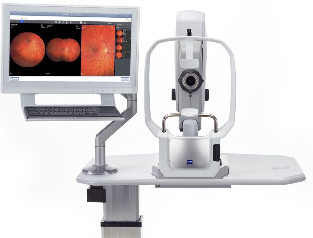
Zeiss CLARUS 500
Examining the entire retina is critical. Some of the most pervasive eye diseases, such as retinal tearing, tumors, and macular degeneration, occur in the far periphery of the eye, making them difficult to detect. Symptoms often do not show up in the early stages and vision may not be affected until there is significant and unrecoverable damage.
The ultra-widefield retina exam is beneficial even if you feel your eyes are healthy. By examining the health of your fundus periodically, your eye doctor can view images from year to year and detect subtle changes in your eyes over time. These changes could go unnoticed and can be the sign of early changes in your eye health.
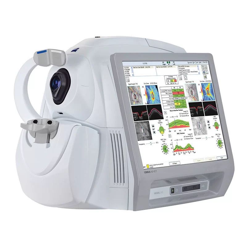
Zeiss OCT 500
An Optical Coherence Tomography scan (commonly referred to as an OCT scan) is the latest advancement in imaging technology. Similar to ultrasound, this diagnostic technique employs light rather than sound waves to achieve higher resolution pictures of the structural layers of the back of the eye. A scanning laser used to analyze the layers of the retina and optic nerve for any signs of eye disease, similar to an CT scan of the eye. It works using light without radiation, and is essential for early diagnosis of glaucoma, macular degeneration and diabetic retinal disease.
With an OCT scan, doctors are provided with color-coded, cross-sectional images of the retina. These detailed images are revolutionizing early detection and treatment of eye conditions such as wet and dry age-related macular degeneration, glaucoma, retinal detachment and diabetic retinopathy. An OCT scan is a noninvasive, painless test. It is performed in less than 5 minutes right in our office.
Zeiss CLARUS 500
Examining the entire retina is critical. Some of the most pervasive eye diseases, such as retinal tearing, tumors, and macular degeneration, occur in the far periphery of the eye, making them difficult to detect. Symptoms often do not show up in the early stages and vision may not be affected until there is significant and unrecoverable damage.
The ultra-widefield retina exam is beneficial even if you feel your eyes are healthy. By examining the health of your fundus periodically, your eye doctor can view images from year to year and detect subtle changes in your eyes over time. These changes could go unnoticed and can be the sign of early changes in your eye health.
Zeiss OCT 500
An Optical Coherence Tomography scan (commonly referred to as an OCT scan) is the latest advancement in imaging technology. Similar to ultrasound, this diagnostic technique employs light rather than sound waves to achieve higher resolution pictures of the structural layers of the back of the eye. A scanning laser used to analyze the layers of the retina and optic nerve for any signs of eye disease, similar to an CT scan of the eye. It works using light without radiation, and is essential for early diagnosis of glaucoma, macular degeneration and diabetic retinal disease. With an OCT scan, doctors are provided with color-coded, cross-sectional images of the retina. These detailed images are revolutionizing early detection and treatment of eye conditions such as wet and dry age-related macular degeneration, glaucoma, retinal detachment and diabetic retinopathy. An OCT scan is a noninvasive, painless test. It is performed in less than 5 minutes right in our office.
LKC RETeval – Electroretinography
Electroretinography is an electrophysiological test of the retina, the layer of the eye which detects light. The electroretinogram (ERG) is to the retina what the electrocardiogram (EKG) is to the heart. Just as an EKG is crucial to diagnosing illness and monitoring the heart’s function, ERG plays a critical role in the care of the eye, and is instrumental in the early detection of retinal dysfunction.
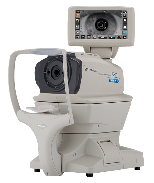
Topcon KR-1 Auto Kerato-Refractometer
The KR-1 Auto Kerato-Refractometer features fully automated operation with an easy-to-use color touch screen, a 360 degree rotatable monitor, and a flexible layout and space saving design. The Rotary Prism measuring system ensures fast, accurate, and repeatable refraction and keratometry measurements.
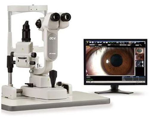
Topcon SL_D4 Imaging System
Our Topcon SL-D701 LED Slit Lamp with Digital Camera and Meibography capabilities combines quality, innovation, and advanced features to deliver exceptional examination and imaging capabilities. It is a versatile and advanced tool for comprehensive eye examinations utilizing precise imaging capabilities. The SL-D701’s advanced features contribute to accurate diagnosis and evaluation of ocular conditions.
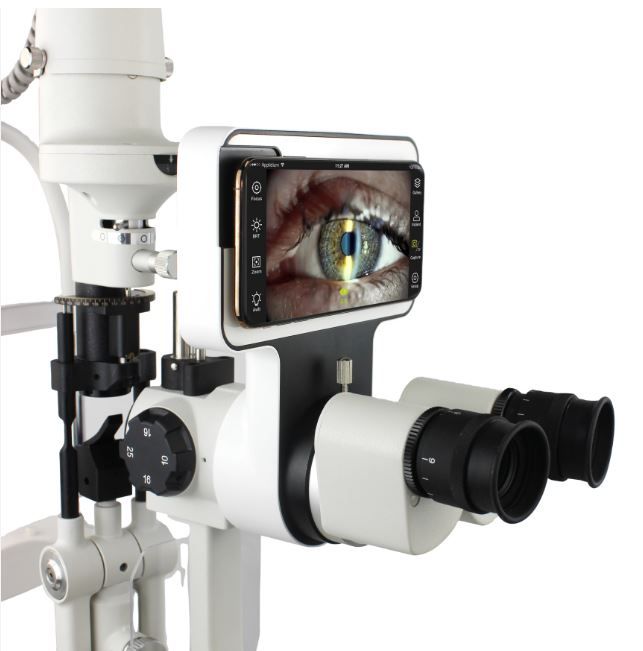
Marco iON Imaging System
Slit lamp imaging is redefined with Marco’s iON anterior segment imaging system by combining a new intra-optics beam splitter/camera adapter with the tremendous computing and imaging power of Apple technology.
Intuitive operation, optimized imaging, and immediate network integration are some trademark qualities of this product.
Humphrey Visual Field
For years, the Humphrey Field Analyzer has brought certainty to glaucoma diagnostics. The HFA3 preserves everything that made its predecessors the gold standard in perimetry—then takes that standard to new heights with innovations that enhance usability to streamline clinic flow. Its uses Liquid Trial lens technology, instantly delivering each patient’s spectacle prescription into the machine, with a touch of a button. Provides the physician with improved confidence in visual field testing with RelEye, instantly reviewing the patient’s eye position at all times.